Modern life presents unprecedented challenges to maintaining proper spinal alignment and postural integrity. Extended periods spent hunched over digital devices, sedentary work environments, and inadequate ergonomic awareness contribute to a widespread epidemic of postural dysfunction affecting millions globally. The human spine, designed for upright mobility and dynamic movement, suffers under the constant stress of forward head posture, rounded shoulders, and compromised spinal curvatures that characterise contemporary postural patterns.
Research consistently demonstrates that targeted corrective exercises can effectively reverse years of postural deterioration when implemented systematically. Unlike passive interventions or expensive equipment-dependent solutions, evidence-based movement protocols offer accessible, cost-effective approaches to restoring optimal spinal alignment. The integration of specific strengthening, mobilisation, and lengthening techniques addresses the root biomechanical causes of postural dysfunction rather than merely treating symptoms.
Understanding the interconnected nature of postural systems enables individuals to develop comprehensive exercise strategies that target multiple dysfunction patterns simultaneously. From cervical spine stabilisation to lumbar-pelvic complex coordination, systematic exercise prescription can dramatically improve postural quality, reduce musculoskeletal pain, and enhance overall functional capacity within weeks of consistent application.
Understanding postural dysfunction and musculoskeletal imbalances
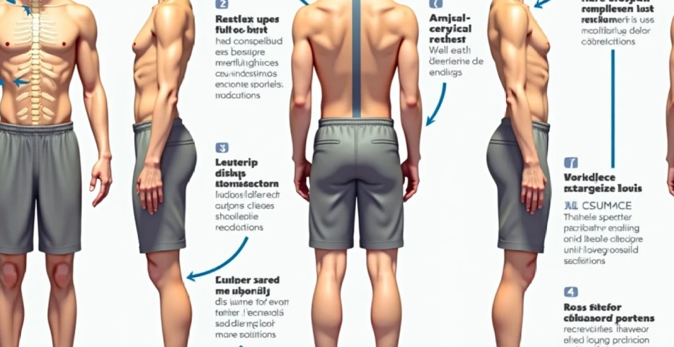
Postural dysfunction represents a complex interplay of muscular imbalances, joint restrictions, and neuromotor control deficits that develop progressively over time. The human body adapts to repetitive positioning through tissue remodelling, creating compensatory patterns that initially maintain function but ultimately compromise structural integrity. These adaptations manifest as predictable syndrome patterns that healthcare professionals recognise as upper crossed syndrome, lower crossed syndrome, and layer syndrome complexes.
Modern postural analysis reveals that gravity-induced stress patterns create systematic weakening of posterior chain muscles whilst simultaneously tightening anterior structures. This imbalance disrupts the natural reciprocal relationship between agonist and antagonist muscle groups, leading to altered joint mechanics and compensatory movement strategies. The cervical spine loses its natural lordotic curve, the thoracic spine develops excessive kyphosis, and the lumbar region may exhibit either hyperlordosis or flattening depending on individual compensation patterns.
Forward head posture and cervical lordosis alterations
Forward head posture represents one of the most prevalent postural dysfunctions in contemporary society, affecting an estimated 66% of office workers and 85% of frequent technology users. This condition develops when the cranium migrates anteriorly relative to the shoulders, creating a cascade of compensatory adaptations throughout the cervical and upper thoracic regions. The suboccipital muscles become hyperactive whilst deep cervical flexors weaken significantly, creating an imbalance that perpetuates the forward head position.
Biomechanical analysis reveals that every inch of forward head migration increases the effective weight of the head by approximately 10-12 pounds, exponentially increasing stress on cervical structures. This increased loading pattern leads to accelerated degenerative changes, including cervical disc compression, facet joint irritation, and myofascial trigger point development. The altered cervical lordosis disrupts normal shock absorption mechanisms, transferring excessive forces to surrounding tissues and creating the characteristic pain patterns associated with cervical dysfunction.
Thoracic kyphosis and upper crossed syndrome patterns
Excessive thoracic kyphosis, commonly termed “hunchback posture,” develops through prolonged flexion positioning that characterises modern work environments. This postural deviation involves increased curvature of the upper back, accompanied by protracted shoulders and internally rotated humeri. The pectoralis minor and major muscles adaptively shorten whilst the middle and lower trapezius, rhomboids, and posterior deltoids become lengthened and weakened.
Upper crossed syndrome, as described by Vladimir Janda, represents the classic muscle imbalance pattern associated with thoracic kyphosis. Tight upper trapezius, levator scapulae, sternocleidomastoid, and pectoral muscles create reciprocal inhibition of their antagonists, establishing a self-perpetuating cycle of dysfunction. This pattern not only affects spinal alignment but also compromises respiratory mechanics, reducing lung capacity and contributing to shallow breathing patterns that further exacerbate postural dysfunction.
Anterior pelvic tilt and lower crossed syndrome manifestations
Anterior pelvic tilt occurs when the pelvis rotates forward, increasing lumbar lordosis and creating characteristic “sway back” posture. This dysfunction primarily results from hip flexor tightness combined with gluteal weakness, creating an imbalance that tilts the pelvis anteriorly and increases stress on lumbar structures. Prolonged sitting positions contribute significantly to this pattern by maintaining hip flexors in shortened positions whilst simultaneously inhibiting gluteal activation.
Lower crossed syndrome encompasses the reciprocal relationship between tight hip flexors and erector spinae muscles with weak abdominals and gluteals. This imbalance pattern creates compensatory lumbar extension, increased disc pressure, and altered load distribution throughout the lumbar spine. The resulting biomechanical dysfunction contributes to lumbar disc degeneration, facet joint arthropathy, and chronic low back pain syndromes that affect millions globally.
Scapular protraction and rounded shoulder mechanics
Scapular protraction involves forward displacement of the shoulder blades, creating the characteristic rounded shoulder appearance common in desk workers and smartphone users. This positioning results from serratus anterior weakness combined with middle trapezius and rhomboid lengthening, disrupting normal scapulohumeral rhythm and glenohumeral joint mechanics. The altered scapular position compromises rotator cuff function and contributes to impingement syndromes.
The biomechanical consequences of scapular protraction extend beyond aesthetic concerns, significantly impacting upper extremity function and cervical spine mechanics. Protracted scapulae reduce subacromial space, increasing impingement risk during overhead activities. Additionally, the altered scapular position affects cervical spine alignment through fascial connections, contributing to neck pain and headache patterns commonly observed in individuals with poor upper body posture.
Evidence-based cervical and thoracic spine strengthening protocols
Cervical and thoracic spine strengthening requires targeted activation of deep stabilising muscles whilst simultaneously addressing mobility restrictions that limit normal movement patterns. Research demonstrates that specific motor control training produces superior outcomes compared to general strengthening approaches, particularly for addressing forward head posture and cervical dysfunction. The integration of endurance-based strengthening with precise movement quality ensures sustainable postural improvements.
Progressive loading principles guide effective cervical and thoracic exercise prescription, beginning with low-threshold activation exercises before advancing to functional integration patterns. This approach respects tissue healing timelines whilst systematically building the strength and endurance necessary for maintaining improved postural alignment throughout daily activities. The emphasis on quality over quantity ensures proper movement patterns become established before increasing exercise intensity or complexity.
Deep cervical flexor activation using Cranio-Cervical flexion test techniques
Deep cervical flexor strengthening represents the cornerstone of cervical spine rehabilitation, targeting the longus colli and longus capitis muscles that provide essential cervical stability. The cranio-cervical flexion test, originally developed as an assessment tool, serves as an excellent therapeutic exercise when applied progressively. This technique involves subtle chin retraction movements performed in supine position, activating deep cervical flexors whilst minimising superficial muscle substitution.
Proper technique involves lying supine with knees bent, performing gentle chin tucks whilst maintaining eye level horizontal. The movement should create a slight lengthening sensation at the base of the skull without engaging superficial neck flexors. Progressive holds of 10-30 seconds repeated 5-10 times establish endurance capacity essential for maintaining cervical alignment during daily activities. Advanced progressions include adding resistance through manual pressure or performing the exercise in various positions to challenge stability requirements.
Rhomboid and middle trapezius strengthening through wall angel progressions
Wall angel exercises provide an excellent foundation for retraining scapular retraction and thoracic extension patterns whilst offering external support for proper alignment. This exercise targets the middle trapezius, rhomboids, and posterior deltoids whilst simultaneously stretching tight pectoral muscles. The wall provides tactile feedback that helps individuals learn proper scapular positioning and movement patterns essential for postural correction.
Initial wall angel technique involves standing with back against wall, arms positioned in “goal post” formation with elbows and wrists maintaining wall contact. Slow sliding movements up and down the wall challenge scapular stability whilst promoting thoracic mobility. Progression involves increasing range of motion, adding isometric holds, or performing the exercise without wall support to enhance proprioceptive demands. Advanced variations include single-arm patterns or resistance band additions to increase strengthening challenges.
Thoracic extension mobilisation via foam roller and towel roll methods
Thoracic extension mobilisation addresses the mobility restrictions that commonly accompany increased kyphosis and rounded shoulder postures. Foam roller techniques provide targeted pressure to thoracic segments whilst facilitating extension movement patterns essential for postural restoration. The combination of passive positioning with active movement creates optimal conditions for tissue remodelling and joint mobility restoration.
Proper foam roller technique involves positioning the roller horizontally across the thoracic spine at various levels, supporting the head with interlaced fingers whilst gently extending over the roller. Movement should be controlled and gradual, avoiding excessive pressure that might cause protective muscle guarding. Towel roll alternatives provide gentler mobilisation suitable for individuals with acute sensitivity or those beginning mobility restoration programmes. Sessions of 2-3 minutes per spinal level, performed daily, typically produce noticeable improvements within 1-2 weeks.
Levator scapulae and upper trapezius lengthening protocols
Systematic lengthening of hypertonic neck and shoulder muscles represents a critical component of comprehensive postural rehabilitation. The levator scapulae and upper trapezius frequently develop adaptive shortening in response to forward head posture and elevated stress levels. Effective stretching protocols must address both the mechanical and neurophysiological aspects of muscle tension to achieve lasting length changes.
Targeted stretching techniques for the levator scapulae involve lateral cervical flexion combined with slight rotation, holding the stretched position for 30-60 seconds. Upper trapezius stretching utilises similar positioning with emphasis on lateral flexion and slight cervical flexion. The addition of gentle overpressure through the contralateral hand enhances stretch effectiveness whilst maintaining safety. Frequency of 3-5 repetitions, 2-3 times daily, produces optimal results when combined with strengthening exercises for antagonist muscles.
Lumbar-pelvic complex stabilisation and hip flexor management
The lumbar-pelvic complex functions as the body’s centre of gravity and primary force transmission hub, requiring optimal stability and mobility for efficient movement patterns. Dysfunction in this region creates compensatory adaptations throughout the kinetic chain, contributing to both spinal pathology and peripheral joint problems. Effective stabilisation strategies must address core endurance, hip mobility, and neuromuscular control simultaneously to achieve comprehensive functional restoration.
Modern research emphasises the importance of coordinated muscle activation patterns rather than isolated strengthening approaches for lumbar-pelvic stability. The integration of deep stabilising muscles including the diaphragm, pelvic floor, multifidus, and transverse abdominis creates an internal cylinder of support that maintains spinal alignment during dynamic activities. This approach produces superior functional outcomes compared to traditional strengthening methods focused on superficial muscle groups.
Hip flexor management represents a crucial component of lumbar-pelvic rehabilitation, as chronic tightness in these muscles directly contributes to anterior pelvic tilt and lumbar hyperlordosis. The psoas major, iliacus, and rectus femoris require systematic lengthening combined with antagonist strengthening to restore balanced pelvic positioning. Progressive stretching protocols must be carefully implemented to avoid aggravating existing lumbar pathology whilst effectively addressing hip flexor restrictions.
Core stabilisation exercises should progress from static holds to dynamic challenges that replicate functional movement demands. Beginning with basic transverse abdominis activation and progressing to complex movement patterns ensures proper motor learning and strength development. The bird-dog exercise exemplifies this progression, starting with simple arm or leg lifts before advancing to opposite limb extensions that challenge spinal stability. Planking variations provide additional challenges for developing core endurance and stability in various planes of movement.
Gluteal strengthening represents another essential component of lumbar-pelvic rehabilitation, as these muscles provide critical hip stability and pelvic alignment control. Weak gluteal muscles contribute to anterior pelvic tilt, increased lumbar lordosis, and compensatory movement patterns that stress spinal structures. Progressive strengthening protocols beginning with basic gluteal activation exercises and advancing to functional movement patterns ensure comprehensive rehabilitation of the posterior chain muscle groups.
Research demonstrates that individuals with chronic low back pain exhibit significantly reduced gluteal activation during functional activities, highlighting the importance of targeted strengthening protocols for these critical stabilising muscles.
Hip flexor stretching techniques must be performed with careful attention to lumbar spine positioning to maximise effectiveness whilst avoiding symptom aggravation. The modified Thomas test position provides an excellent framework for systematic hip flexor lengthening, allowing targeted stretching of specific muscle groups. Kneeling hip flexor stretches offer dynamic alternatives that can be easily incorporated into daily routines, providing consistent stimulus for tissue adaptation and length restoration.
Workplace ergonomic integration and movement pattern corrections
Workplace environments significantly influence postural development, with poorly designed workstations contributing to accelerated postural dysfunction development. The integration of ergonomic principles with corrective exercise strategies creates comprehensive approaches that address both causative factors and symptomatic presentations. Modern workplace interventions must consider technological demands whilst promoting movement variability and postural awareness throughout the working day.
Movement pattern corrections require systematic retraining of habitual positioning and movement strategies that contribute to postural dysfunction. The human nervous system readily adapts to repetitive patterns, making conscious retraining essential for breaking destructive postural habits. This process involves developing proprioceptive awareness, implementing movement cues, and creating environmental supports that facilitate better postural choices throughout daily activities.
Workstation setup following HSE display screen equipment regulations
Proper workstation ergonomics forms the foundation of postural health in office environments, requiring systematic attention to monitor height, keyboard positioning, and chair adjustments. The Health and Safety Executive Display Screen Equipment regulations provide evidence-based guidelines for optimising workstation design to minimise postural stress. Monitor positioning at eye level reduces cervical flexion demands, whilst proper keyboard height maintains neutral wrist positioning and reduces upper extremity tension.
Chair selection and adjustment significantly impacts spinal alignment and muscle activation patterns during prolonged sitting. Appropriate lumbar support maintains the natural lordotic curve whilst reducing disc pressure and muscle fatigue. Armrest positioning should support the arms without elevating the shoulders, reducing upper trapezius tension and promoting relaxed shoulder positioning. Regular workstation assessments ensure continued optimisation as individual needs and equipment wear patterns evolve over time.
Pomodoro technique movement breaks and Micro-Exercise implementation
The Pomodoro Technique’s structured work-break cycles provide excellent frameworks for implementing regular movement interventions throughout the workday. Strategic placement of micro-exercises during natural break periods prevents tissue stiffness accumulation whilst promoting circulation and muscle activation. These brief interventions, lasting 1-3 minutes, can significantly impact postural health when implemented consistently over time.
Effective micro-exercises target the primary dysfunction patterns associated with prolonged sitting, including cervical retraction, thoracic extension, and hip flexor stretching. Simple exercises such as doorway chest stretches, seated spinal twists, and standing hip flexor stretches can be performed in office environments without requiring special equipment or clothing changes. The key lies in consistency and progressive integration rather than exercise intensity or complexity.
Alexander technique principles for daily movement awareness
Alexander Technique principles emphasise conscious awareness and control of movement patterns, providing valuable frameworks for postural re-education. The concept of “use affects function” highlights how habitual movement patterns influence structural adaptation and pain development. Integration of these principles into daily activities creates opportunities for continuous postural improvement without requiring dedicated exercise time.
Practical application involves developing awareness of head-neck-spine relationships during routine activities such as walking, sitting, and reaching. The principle of “lengthening and widening” encourages spinal decompression and optimal alignment during movement transitions. Regular check-ins throughout the day help establish new habit patterns that support improved posture naturally and effortlessly.
Progressive exercise prescription and biomechanical assessment markers
Effective postural rehabilitation requires systematic progression from basic movement re-education to complex functional integration patterns. Progressive exercise prescription ensures appropriate challenge levels whilst respecting tissue healing timelines and individual adaptation rates. The integration of biomechanical assessment markers provides objective measures for monitoring progress and adjusting exercise protocols based on individual response patterns
that support improved postural outcomes. Objective measurement protocols enable practitioners to track changes in spinal alignment, muscle activation patterns, and functional movement quality throughout the rehabilitation process.
Standardised assessment tools provide reliable frameworks for quantifying postural improvements and identifying areas requiring continued intervention. Photogrammetric analysis offers non-invasive methods for documenting changes in head position, shoulder alignment, and spinal curvatures over time. These measurements complement subjective pain assessments and functional questionnaires to create comprehensive pictures of postural rehabilitation progress.
Exercise progression follows established motor learning principles, beginning with conscious competence phases before transitioning to automatic movement integration. Initial phases emphasise movement quality and proprioceptive awareness, gradually increasing complexity and functional relevance as movement patterns become established. The integration of feedback mechanisms, including visual, tactile, and proprioceptive cues, accelerates learning and ensures sustainable postural improvements.
Biomechanical markers for assessing postural improvement include cervical lordosis restoration, thoracic kyphosis normalisation, and lumbar curve optimisation measured through clinical photography or postural grid analysis. Functional assessments evaluate movement quality during activities of daily living, including sit-to-stand transitions, reaching patterns, and gait mechanics. These combined measures provide comprehensive evaluation frameworks that guide exercise prescription and track rehabilitation outcomes effectively.
Progressive exercise prescription should challenge the individual without overwhelming their adaptive capacity, maintaining the delicate balance between stimulus and recovery that promotes optimal tissue remodelling and neuromotor learning.
Resistance training progressions incorporate traditional strengthening principles whilst addressing specific postural dysfunction patterns. Beginning with bodyweight exercises and progressing to external resistance ensures adequate strength development for maintaining improved postural alignment against gravitational forces. The timing of progression depends on individual adaptation rates, with typical advancement criteria including pain-free movement execution and demonstrated movement quality maintenance under increased demands.
Long-term maintenance strategies emphasise habit formation and environmental modification rather than relying solely on structured exercise compliance. The development of postural awareness cues and movement reminders creates sustainable behavioural changes that persist beyond formal rehabilitation periods. Regular reassessment ensures continued progress and identifies emerging dysfunction patterns before they become symptomatic, supporting lifelong postural health maintenance.
Exercise prescription individualisation considers personal factors including age, fitness level, existing pathology, and lifestyle demands. Older adults may require longer adaptation periods and gentler progression rates, whilst athletic populations can typically tolerate more aggressive advancement schedules. The presence of acute pathology necessitates modified approaches that respect tissue healing constraints whilst promoting optimal recovery outcomes.
Technology integration enhances exercise prescription through wearable devices that monitor posture throughout daily activities, providing real-time feedback and objective progress tracking. Smartphone applications offer convenient exercise reminders and instructional videos that support home-based rehabilitation programmes. These tools complement professional guidance whilst empowering individuals to take active roles in their postural rehabilitation journeys.
Multidisciplinary approaches incorporating physiotherapy, occupational therapy, and ergonomic consultation produce superior outcomes compared to single-intervention strategies. The combination of manual therapy techniques with corrective exercise addresses both structural restrictions and movement pattern dysfunctions comprehensively. Regular communication between healthcare providers ensures coordinated care that maximises rehabilitation efficiency and minimises treatment redundancy.
Success metrics extend beyond simple pain reduction to include functional capacity improvements, postural alignment restoration, and quality of life enhancements. Validated outcome measures such as the Neck Disability Index, Oswestry Disability Index, and postural stability assessments provide standardised frameworks for documenting rehabilitation effectiveness. These comprehensive evaluation approaches demonstrate the multifaceted benefits of systematic postural rehabilitation programmes implemented through evidence-based exercise prescription protocols.