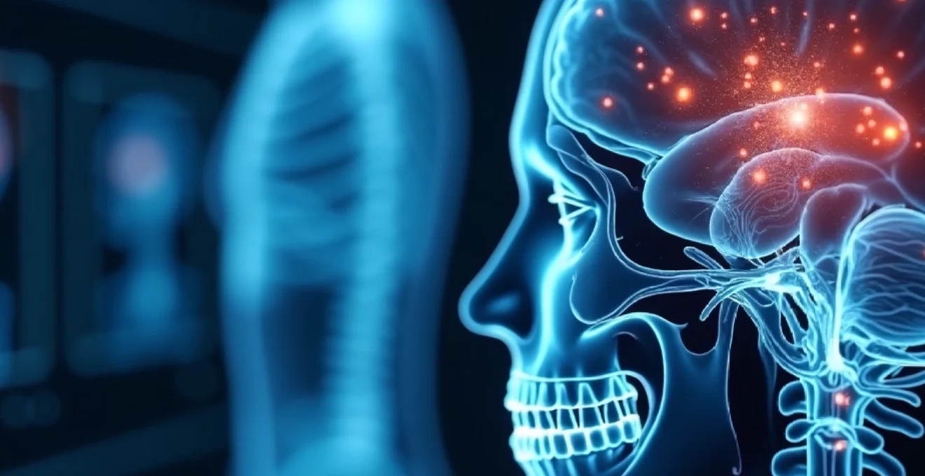
Medical imaging has revolutionised the landscape of healthcare by enabling clinicians to identify diseases before symptoms manifest, fundamentally transforming patient outcomes and survival rates. Through sophisticated technologies ranging from computed tomography to molecular imaging, healthcare professionals can now detect cellular changes, tissue abnormalities, and physiological dysfunction at their earliest stages. Early detection through imaging modalities has demonstrated remarkable success in reducing mortality rates across multiple disease categories, with breast cancer screening programmes showing up to 40% reduction in deaths when mammography is implemented systematically. The ability to visualise internal structures without invasive procedures has made screening programmes accessible to larger populations, creating opportunities for preventive intervention rather than reactive treatment.
Radiological screening technologies for asymptomatic disease detection
Contemporary radiological screening represents a paradigm shift from symptomatic diagnosis to proactive health monitoring, utilising advanced imaging technologies to identify pathological changes in asymptomatic individuals. These sophisticated screening protocols have transformed public health outcomes by detecting diseases at stages when therapeutic interventions are most effective and less invasive.
Low-dose CT scanning for lung cancer detection in High-Risk populations
Low-dose computed tomography (LDCT) has emerged as the gold standard for lung cancer screening, particularly amongst high-risk populations including long-term smokers and individuals with occupational exposure to carcinogens. This technology reduces radiation exposure by approximately 90% compared to standard CT protocols whilst maintaining diagnostic accuracy for detecting small pulmonary nodules. The National Lung Screening Trial demonstrated that LDCT screening reduces lung cancer mortality by 20% in high-risk populations, making it one of the most effective cancer screening modalities available.
The protocol involves annual screening for individuals aged 50-80 years with a 20 pack-year smoking history, identifying nodules as small as 4 millimetres in diameter. Advanced computer-aided detection algorithms enhance radiologist interpretation, reducing false-positive rates whilst maintaining sensitivity for malignant lesions. Implementation challenges include managing incidental findings and ensuring appropriate follow-up protocols for detected abnormalities.
Digital mammography and tomosynthesis for breast cancer screening protocols
Digital breast tomosynthesis, often referred to as 3D mammography, has significantly improved breast cancer detection rates whilst reducing false-positive results compared to conventional 2D mammography. This technology creates multiple thin-section images of breast tissue, allowing radiologists to examine overlapping structures that might obscure small cancers in traditional mammographic images. Studies indicate that tomosynthesis increases cancer detection rates by 41% compared to digital mammography alone.
The integration of artificial intelligence algorithms in mammographic interpretation has further enhanced diagnostic accuracy, with some systems demonstrating performance comparable to expert radiologists. Screening protocols typically commence at age 40-50 depending upon risk factors and national guidelines, with annual or biennial intervals. Dense breast tissue presents particular challenges, as it can mask small tumours and increase the likelihood of false-negative results, prompting supplementary screening with ultrasound or MRI in high-risk patients.
Colonoscopy virtual reality and CT colonography for colorectal malignancy detection
CT colonography, commonly known as virtual colonoscopy, offers a minimally invasive alternative to conventional colonoscopy for colorectal cancer screening. This technique utilises multi-detector CT technology to generate high-resolution images of the colon and rectum, enabling detection of polyps and early-stage cancers without requiring sedation or invasive instrumentation. Virtual colonoscopy demonstrates sensitivity rates of 90% for polyps larger than 10 millimetres, comparable to optical colonoscopy for clinically significant lesions.
The procedure requires bowel preparation similar to conventional colonoscopy but eliminates risks associated with sedation and perforation. Advanced post-processing software creates three-dimensional reconstructions of the colon, allowing radiologists to navigate virtually through the bowel lumen. Patient acceptance rates are higher for CT colonography due to reduced discomfort and shorter examination times, potentially increasing screening participation rates in target populations.
Cardiac CT angiography for coronary artery disease assessment
Coronary CT angiography (CCTA) has transformed cardiac risk assessment by providing non-invasive visualisation of coronary arteries and quantification of atherosclerotic plaque burden. This technology enables detection of subclinical coronary artery disease before symptom onset, facilitating early intervention with lifestyle modifications and preventive therapies. CCTA demonstrates negative predictive values exceeding 95% for excluding significant coronary artery disease, making it particularly valuable for ruling out cardiac pathology in low-to-intermediate risk patients.
Advanced plaque analysis algorithms can differentiate between calcified, non-calcified, and mixed plaque compositions, providing insights into plaque vulnerability and rupture risk. The coronary artery calcium score derived from CT imaging correlates strongly with cardiovascular event risk, enabling more precise risk stratification than traditional scoring systems alone. Radiation exposure considerations have led to development of ultra-low dose protocols that maintain diagnostic quality whilst minimising patient radiation burden.
Bone densitometry DEXA scanning for osteoporosis risk stratification
Dual-energy X-ray absorptiometry (DEXA) scanning represents the gold standard for osteoporosis screening and fracture risk assessment, utilising low-energy X-rays to measure bone mineral density at key skeletal sites. This technology enables detection of osteoporosis before fractures occur, facilitating early intervention with bone-protective therapies. DEXA scanning demonstrates excellent precision and reproducibility, with measurement errors typically less than 2% for spine and hip assessments.
The World Health Organisation criteria define osteoporosis using T-scores derived from DEXA measurements, with values below -2.5 standard deviations indicating significant bone loss requiring treatment. Recent advances include trabecular bone score analysis, which assesses bone microarchitecture quality beyond density measurements. Screening protocols typically recommend DEXA scanning for women over 65 years and men over 70 years, with earlier screening for individuals with risk factors such as steroid use or family history of osteoporotic fractures.
Advanced MRI techniques in Pre-Clinical disease identification
Magnetic resonance imaging has evolved far beyond anatomical visualisation to encompass functional and molecular imaging capabilities that detect disease processes at their earliest stages. These advanced MRI techniques exploit physiological parameters such as tissue perfusion, cellular metabolism, and molecular diffusion to identify pathological changes before structural abnormalities become apparent on conventional imaging.
Diffusion-weighted imaging for early stroke detection and brain tumour characterisation
Diffusion-weighted imaging (DWI) exploits the random motion of water molecules in biological tissues to detect acute ischaemic changes within minutes of stroke onset, significantly earlier than conventional MRI sequences. This technique measures apparent diffusion coefficient values, which decrease rapidly in areas of cytotoxic oedema following arterial occlusion. DWI demonstrates sensitivity rates exceeding 95% for acute stroke detection within the first hour of symptom onset, enabling rapid treatment decisions for thrombolytic therapy.
In oncological applications, DWI provides insights into tumour cellularity and treatment response by measuring water diffusion restriction within malignant tissues. High-grade tumours typically demonstrate significantly reduced diffusion coefficients compared to low-grade lesions, enabling non-invasive tumour grading. Quantitative analysis of diffusion parameters can predict treatment response before anatomical changes become evident, facilitating earlier therapy modifications in cases of treatment failure.
Functional MRI biomarkers in alzheimer’s disease and dementia progression
Functional MRI techniques, including blood-oxygen-level-dependent (BOLD) imaging and arterial spin labelling, detect subtle brain function changes years before clinical symptoms of dementia manifest. These methodologies measure brain activation patterns, connectivity networks, and cerebral perfusion alterations associated with neurodegenerative processes. Resting-state functional connectivity MRI can identify disruptions in default mode networks that occur in preclinical Alzheimer’s disease stages.
Quantitative perfusion imaging reveals regional hypoperfusion patterns that correlate with cognitive decline risk, enabling early identification of individuals likely to develop dementia. Advanced post-processing techniques measure network connectivity metrics and graph theory parameters that reflect brain organisational efficiency. Longitudinal studies demonstrate that functional MRI biomarkers can predict cognitive decline up to five years before clinical diagnosis, providing opportunities for early therapeutic intervention and lifestyle modifications.
Cardiac MRI perfusion studies for myocardial ischaemia detection
Stress perfusion cardiac MRI utilises pharmacological vasodilation to assess myocardial blood flow and detect coronary artery disease before symptom development or wall motion abnormalities occur. This technique combines high spatial resolution with excellent temporal resolution to identify perfusion defects during peak hyperaemia induced by adenosine or regadenoson administration. Cardiac MRI perfusion demonstrates diagnostic accuracy comparable to nuclear perfusion imaging whilst avoiding ionising radiation exposure.
Quantitative perfusion analysis measures absolute myocardial blood flow values rather than relative perfusion differences, enabling detection of balanced three-vessel disease that might be missed by relative perfusion techniques. T1 mapping sequences assess myocardial tissue characteristics, identifying diffuse fibrosis and infiltrative cardiomyopathies at subclinical stages. Parametric mapping techniques provide pixel-by-pixel quantification of tissue properties, enabling detection of subtle myocardial abnormalities before global function deterioration becomes apparent.
MR spectroscopy applications in metabolic disorder diagnosis
Magnetic resonance spectroscopy (MRS) provides non-invasive assessment of tissue metabolism by measuring concentrations of specific metabolites within defined anatomical regions. This technique enables detection of metabolic abnormalities before structural changes occur, particularly valuable in neurological and hepatic disorders. Proton MRS can quantify key brain metabolites including N-acetylaspartate, choline, creatine, and lactate, providing insights into neuronal integrity and metabolic function.
Hepatic MRS applications include non-invasive quantification of liver fat content, enabling early detection of non-alcoholic fatty liver disease before progression to steatohepatitis or cirrhosis. Phosphorus-31 MRS assesses cellular energy metabolism by measuring adenosine triphosphate and phosphocreatine levels, particularly valuable in cardiac and skeletal muscle disorders. Metabolic fingerprinting approaches utilise pattern recognition algorithms to identify disease-specific metabolite profiles that may precede conventional diagnostic markers.
Molecular imaging and nuclear medicine in disease biomarker detection
Nuclear medicine techniques harness the power of radiotracer technology to visualise biological processes at the molecular and cellular level, providing unparalleled sensitivity for detecting disease in its earliest stages. These methodologies combine sophisticated radiopharmaceutical chemistry with advanced imaging hardware to identify pathophysiological changes that occur before anatomical abnormalities become apparent through conventional imaging modalities.
PET-CT fluorodeoxyglucose imaging for oncological staging and metastasis detection
Fluorodeoxyglucose (FDG) positron emission tomography combined with computed tomography represents one of the most powerful tools for cancer detection and staging, exploiting increased glucose metabolism characteristic of malignant cells. This technique detects tumours as small as 5-10 millimetres whilst simultaneously assessing metabolic activity levels that correlate with tumour aggressiveness and treatment response. FDG-PET demonstrates sensitivity rates exceeding 90% for detecting most solid tumours, with particularly high accuracy in lung cancer, lymphoma, and melanoma.
The integration of PET and CT technologies provides both functional and anatomical information in a single examination, enabling precise localisation of metabolically active lesions whilst characterising their morphological features. Quantitative analysis using standardised uptake values (SUV) enables objective assessment of tumour metabolism and monitoring of treatment response. Advanced reconstruction algorithms and time-of-flight technology have significantly improved image quality whilst reducing examination times and radiation exposure.
The ability to detect cancer before it becomes clinically apparent has transformed oncological practice, enabling earlier intervention when treatments are most effective and curative potential highest.
SPECT imaging applications in neurological and cardiac pathology assessment
Single photon emission computed tomography (SPECT) imaging utilises gamma-ray emitting radiopharmaceuticals to assess regional organ function and detect pathological processes before structural changes become apparent. In neurological applications, perfusion SPECT with technetium-99m radiopharmaceuticals can identify regional cerebral blood flow abnormalities associated with early dementia, stroke, and epilepsy. These functional changes often precede anatomical alterations by months or years, enabling earlier diagnosis and intervention.
Cardiac SPECT imaging remains the most widely performed nuclear medicine procedure, providing assessment of myocardial perfusion under rest and stress conditions to detect coronary artery disease. The technique demonstrates high diagnostic accuracy for identifying haemodynamically significant coronary stenoses whilst providing prognostic information regarding future cardiac event risk. Recent technological advances including cadmium-zinc-telluride detector systems have significantly improved image quality whilst reducing radiation exposure and examination times.
Radiopharmaceutical tracers for amyloid beta detection in cognitive decline
Amyloid imaging agents such as florbetapir, flutemetamol, and florbetaben enable direct visualisation of amyloid-beta plaque deposition in the living brain, revolutionising Alzheimer’s disease diagnosis and research. These radiopharmaceuticals cross the blood-brain barrier and bind specifically to fibrillar amyloid plaques, providing quantitative assessment of amyloid burden years before clinical symptoms develop. Studies demonstrate that amyloid PET can identify individuals at risk for cognitive decline up to 15 years before symptom onset.
The integration of amyloid imaging with other biomarkers including cerebrospinal fluid analysis and tau PET imaging enables more precise staging of Alzheimer’s disease pathology according to the amyloid-tau-neurodegeneration framework. Visual and quantitative interpretation methods have been standardised to ensure consistent reporting across different imaging centres and reader experience levels. Clinical applications include differential diagnosis of dementia syndromes, clinical trial participant selection, and monitoring of anti-amyloid therapeutic interventions.
Gallium-68 DOTATATE PET scanning for neuroendocrine tumour identification
Gallium-68 DOTATATE PET imaging exploits the overexpression of somatostatin receptors on neuroendocrine tumours to provide highly sensitive and specific detection of these often challenging-to-diagnose malignancies. This radiopharmaceutical demonstrates superior performance compared to conventional imaging modalities, with sensitivity rates exceeding 95% for well-differentiated neuroendocrine tumours. The technique can detect tumours as small as 2-3 millimetres whilst providing whole-body staging information in a single examination.
The high target-to-background ratio achieved with DOTATATE imaging enables detection of small primary tumours and metastatic deposits that may be occult on anatomical imaging. Quantitative analysis using SUV measurements correlates with somatostatin receptor expression density, providing insights into tumour biology and treatment selection. Theranostic applications utilise the same molecular target for both imaging and therapy, with lutetium-177 DOTATATE providing targeted radiotherapy for patients with somatostatin receptor-positive tumours identified through gallium-68 DOTATATE PET imaging.
Ultrasound doppler technology in vascular and organ disease screening
Ultrasound imaging has evolved into a sophisticated diagnostic modality that extends far beyond basic anatomical visualisation to encompass advanced Doppler techniques, elastography, and contrast-enhanced imaging capabilities. The real-time nature of ultrasound examination combined with its non-invasive character and absence of ionising radiation makes it particularly valuable for screening asymptomatic populations and detecting early-stage pathological processes.
Doppler ultrasound technology enables assessment of blood flow patterns and vascular architecture, facilitating early detection of atherosclerotic disease before clinical symptoms develop. Carotid intima-media thickness measurement serves as a surrogate marker for systemic atherosclerosis, with increased thickness values correlating with cardiovascular event risk. Advanced Doppler techniques can detect flow disturbances associated with early arterial stenosis, enabling intervention before complete vascular occlusion occurs.
Shear wave elastography represents a revolutionary advancement in ultrasound technology, providing quantitative assessment of tissue stiffness properties that correlate with pathological changes. This technique enables non-invasive assessment of liver fibrosis, identifying early stages of chronic liver disease before progression to cirrhosis. In breast imaging, elastography enhances
differentiation between malignant and benign lesions with greater accuracy than conventional ultrasound alone. The technique measures tissue elasticity by applying acoustic radiation force impulses, generating shear waves that propagate through tissues at speeds proportional to tissue stiffness.
Contrast-enhanced ultrasound (CEUS) utilises microbubble contrast agents to improve visualisation of organ perfusion and detect pathological processes such as tumours, inflammation, and ischaemia. This technique provides real-time assessment of tissue vascularity without the nephrotoxic risks associated with iodinated contrast agents used in CT imaging. CEUS demonstrates particular utility in liver imaging, where it can characterise focal lesions and detect hepatocellular carcinoma at earlier stages than conventional imaging modalities. Point-of-care ultrasound applications have expanded screening capabilities beyond traditional radiology departments, enabling primary care physicians to perform targeted assessments for conditions such as abdominal aortic aneurysms and deep vein thrombosis.
Artificial intelligence integration in medical imaging diagnostic accuracy
Artificial intelligence algorithms have fundamentally transformed medical imaging interpretation by enhancing diagnostic accuracy, reducing interpretation times, and identifying subtle pathological changes that might escape human detection. Deep learning neural networks trained on vast imaging datasets can now match or exceed expert radiologist performance in specific diagnostic tasks, whilst providing consistent interpretation quality regardless of reader experience or fatigue levels. AI-powered computer-aided detection systems demonstrate particular strength in screening mammography, where they reduce false-negative rates by up to 9.4% whilst maintaining acceptable false-positive rates.
Machine learning algorithms excel at pattern recognition tasks, identifying complex imaging features that correlate with disease presence or progression risk. In chest radiography, AI systems can detect pneumonia, tuberculosis, and lung cancer with sensitivity and specificity rates comparable to experienced radiologists. Advanced neural networks analyse pixel-level image data to generate probability maps highlighting areas of suspected pathology, providing visual guidance to assist human interpretation. Ensemble learning approaches combine multiple AI models to improve diagnostic performance beyond what individual algorithms can achieve, creating robust systems less susceptible to individual model limitations or biases.
Natural language processing capabilities enable AI systems to extract relevant clinical information from patient records, integrating clinical context with imaging findings to provide more accurate diagnoses. These systems can automatically generate structured reports, prioritise urgent cases, and trigger clinical decision support alerts when critical findings are detected. Real-time AI analysis during image acquisition can guide technologists to optimise examination protocols and ensure adequate image quality for diagnostic interpretation. Workflow integration challenges include ensuring seamless embedding of AI tools within existing radiology information systems whilst maintaining appropriate human oversight and clinical responsibility for final diagnostic decisions.
Comparative analysis of imaging modalities for population health screening programmes
Population-based screening programmes require careful consideration of imaging modality selection based on target disease characteristics, population demographics, cost-effectiveness, and infrastructure requirements. Each imaging technique offers distinct advantages and limitations that must be weighed against screening programme objectives and available resources. Successful screening programmes demonstrate clear evidence of mortality benefit, acceptable harm-to-benefit ratios, and sustainable implementation within existing healthcare systems.
Mammography screening programmes represent the most established population-based imaging initiative, with extensive evidence demonstrating mortality reduction in women aged 50-69 years. However, overdiagnosis concerns and false-positive rates have prompted evaluation of risk-stratified screening approaches using additional modalities such as MRI for high-risk women or abbreviated MRI protocols for broader populations. Low-dose CT lung cancer screening shows promise for high-risk smokers but requires sophisticated infrastructure and expertise to manage incidental findings appropriately. Cost-effectiveness analyses must consider not only immediate screening costs but also downstream diagnostic workup, treatment costs, and quality-adjusted life years gained through early detection.
Emerging screening applications include coronary artery calcium scoring for cardiovascular risk assessment, hepatic ultrasound for fatty liver disease detection, and whole-body MRI for cancer screening in high-risk populations. These innovative approaches require robust evidence generation through randomised controlled trials to establish clinical effectiveness before widespread implementation. Population acceptance and participation rates significantly influence screening programme success, with factors such as examination comfort, convenience, and cultural considerations affecting uptake rates. Technological convergence trends suggest future screening programmes may utilise multi-modal imaging approaches, combining complementary techniques to maximise detection sensitivity whilst minimising false-positive results and unnecessary interventions.
The future of medical imaging lies not in replacing human expertise but in augmenting clinical decision-making through intelligent integration of advanced technologies that enhance our ability to detect, characterise, and monitor disease progression at the earliest possible stages.
Quality assurance programmes ensure consistent imaging performance across screening sites through standardised protocols, equipment calibration, and reader training programmes. International guidelines provide frameworks for establishing screening programmes whilst allowing adaptation to local healthcare contexts and resource availability. The integration of artificial intelligence tools promises to enhance screening programme efficiency and accuracy whilst addressing workforce limitations in many healthcare systems. Future developments will likely focus on personalised screening strategies that utilise individual risk factors, genetic information, and previous imaging results to optimise screening intervals and modality selection for each participant.